Diagnostic digital infrared thermography
A risk-free and quick diagnostic method
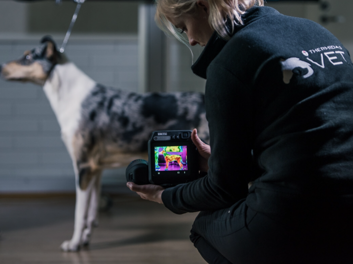
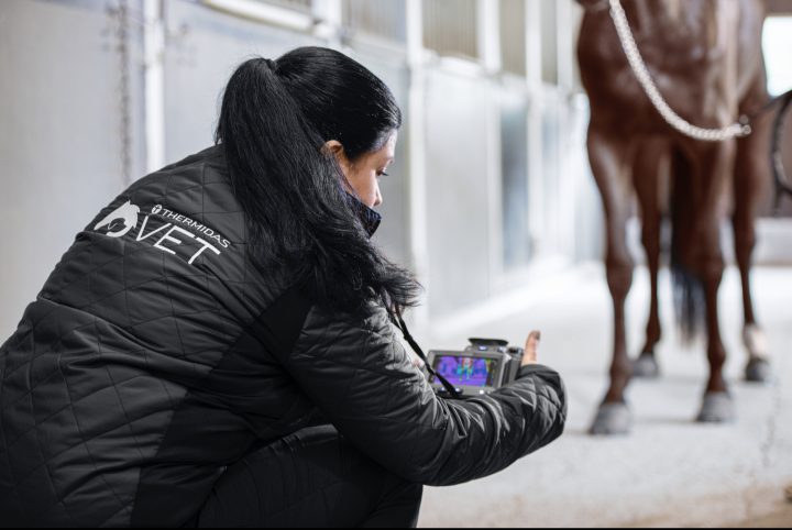
Diagnostic thermal imaging is a comprehensive and risk-free equine and veterinary method for identifying possible injuries and detecting the areas that should be examined further and treated.
Veterinary thermal imaging is done using thermal cameras with especially high IR resolution that can detect temperature variations as low as one-tenth degree Celsius on the animal’s skin surface. We humans can only sense temperature variations that are at least two degrees Celsius. Furthermore, palpation by hand can of course not be used for measuring exact temperature values accurately.
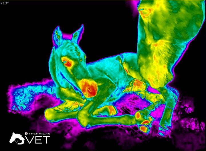
The thermal pattern on the skin of a healthy animal is always symmetrical; there are no differences from side to side. In thermal image analysis, asymmetries that meet the diagnostic criteria are examined in order to locate possible issues quickly and reliably. On its own, thermography may not be enough for reaching a conclusive diagnosis. However, it may critically speed up the detection of injuries and inflammations thus leading to earlier and more accurate treatment. Thermal imaging is a novel supplementary diagnostic method, and its usage requires special expertise. Only trained professionals can take trustworthy thermal images and reliably analyze the results. Diagnosis will always be made by a veterinarian.
Thermography reveals even the tiniest abnormalities. Early detection means quicker treatment, which in turn often translates into lower treatment costs and shorter recovery. Thermal imaging is a superb tool for early diagnostics, because it may enable the detection of possible problems even weeks before any clinical signs appear.
Thermal imaging can be used to quickly and reliably detect changes in
- muscular,
- vascular,
- skeletal, and
- nervous systems.
Thermal imaging graphically shows the animal’s physiological condition by mapping skin surface temperature. The thermal patterns on both sides of a healthy animal are symmetrical (controlled by the sympathetic nervous system). Abnormalities can be detected from differences between skin surface temperature patterns on either side.
Clinical applications include
- Early injury detection before the appearance of clinical signs. (For instance, since a horse is prey, it may not show pain. Thermography can also be used to comprehensively examine animals in pain as well as rescue animals, since the animal is not touched during the examination.)
- Localizing problem areas for further examinations and appropriate treatment.
- Determining the size of the wound/injury.
- Evaluating treatment impact and monitoring healing.
- An animal-friendly diagnostic tool, since it is not based on identifying pain reaction.
The benefits of thermography
- A safe and non-invasive examination.
- Examination can be done from a distance. A risk-free and non-contact diagnostic method even for animals in pain.
- Quick, objective and reliable measurement of local temperature changes and the identification of possible asymmetries.
- Pleasant for the animal, not based on pain reaction identification.
- No sedation (no withdrawal period).
- No fur clipping.
- No exposure to radiation.
- Safe also for pregnant and lactating animals, newborns, young and very old animals.
- Immediate results in graphic, visual form.